It covers the following examination methods:
rtg diagnostics
ultrasound examination of high frequency probes
CT scan
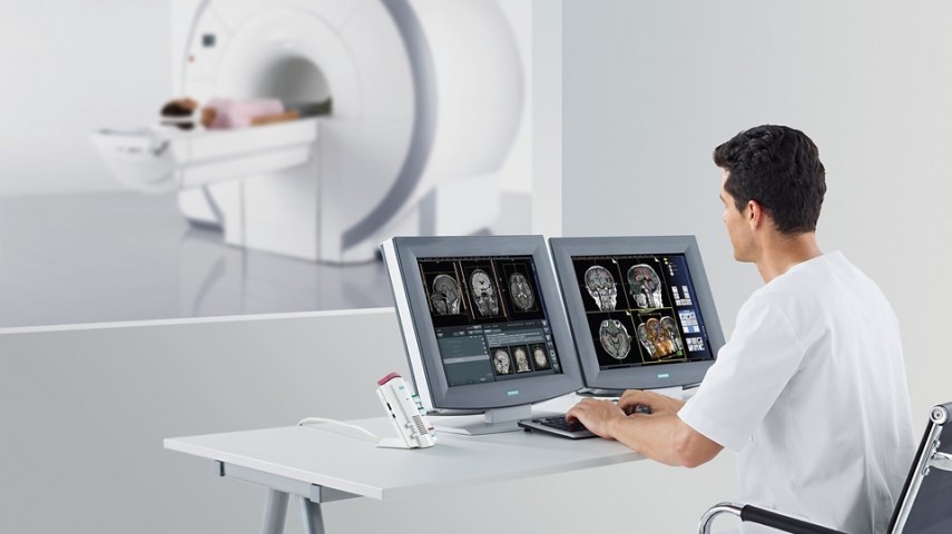
MRI examination of joints, bones, muscles, spine
EMNG-testing of nerve conduction and assessment of intensity and site of damage
Which method will be used depends on the problem to be solved, usually a combination of two or more examination techniques is required.
-
RTG Radiography - is the oldest and most commonly used diagnostic method of body imaging.
Bone-joint X-ray is used for:
Detection of bone fractures and joint injuries and other bone injuries,
View fractured bone sections after various fracture treatments, and monitor fracture healing,
Display of bone changes in the joints in inflammatory joint disease,
Display of bone anomalies,
Presentation of bone changes in metabolic diseases (eg osteoporosis), inflammation, and bone growth disorders,
Assistance in detecting and diagnosing bone tumors,
Detection of foreign bodies visible on the rtg. shots in the soft tissues around or in the bone itself,
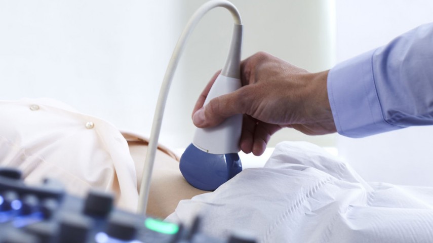
Musculoskeletal ultrasound diagnostics (ultrasound)
It is a radiological method that has made great progress and has gained in popularity and breadth of use in the diagnosis of primarily muscle, tendon, ligaments, joints, and other soft body structures. The advantage of ultrasound over other methods is that it is cheaper, comfortable, has no radiation, can be repeated numerous times as needed, joints, ligaments can be displayed as well as blood flow through the blood vessels by color Doppler and exclude or confirm the existence of thrombosis.
Ultrasound imaging of the musculoskeletal system is commonly used to diagnose:
tendon injuries such as injury to the tendons of the rotator cuff or Achilles tendon,
muscle abnormalities, such as muscle rupture or soft tissue formation,
bleeding and other fluid buildup in muscles, bursas or joints, or blood vessel thrombosis,
less benign and malignant soft tissue tumors,
early changes in inflammatory joint disease (eg rheumatoid arthritis).
The ultrasound waves hardly penetrate the bone, so it shows only the outer contour of the bone. We use rtg and CT to show the bones, and primarily MRI to show the joints. Also, ultrasound-controlled interventions such as cyst, effusion, hematoma or tumor puncture, aspiration, intra-articular infiltration, or installation can be done.
Magnetic resonance imaging (MR) is, due to its high contrast resolution, the method of choice in the diagnosis of musculoskeletal system (trauma, sports injuries and overuse syndromes, inflammatory diseases, changes in degenerative joint disease and tumors) and the only non-invasive diagnostic method that can present within the joint structures (articular cartilage, menisci, and ligaments).
Sometimes it is necessary to further increase the sensitivity of this imaging method in intra-articular defects and to provide a contrast agent to the joint, called direct MR arthrography, which involves invasive diagnostic testing.
-
CT-computerized tomography is nowadays less commonly used in the use of diagnostics of musculoskeletal problems, in addition to radiographs, ultrasound and MRI, primarily due to radiation accompanying the use of CT, but in some cases it is necessary.
Electromyoneurography (EMNG) is a diagnostic method that tests the electrical activity of muscles and nerves with the ability to determine the level, extent and extent of the lesion, usually examining the conductivity of peripheral nerves, motor and / or sensory, first.
The results obtained I can point to various damages of the peripheral nervous and muscular system, that is, neuromuscular disease. In most cases, the optimal time for an EMNG examination is three weeks after the onset of symptoms, due to the development of an electrophysiological picture of the disease or injury.
It works with:
Radiculopathies - EMNG provides information on the level and extent of damage, most commonly of lumbosacral or cervical nerve roots (one of the basic criteria for setting an indication for surgical treatment and evaluating its success).
Focal neuropathies - traumatic nerve damage, nerve compression over a long period of time (eg sleep paralysis n radials), entrapment of a nerve in a fibrous or bone canal or between fibrous bands and bones (eg Carpal tunnel sy), infarction, tumor or nerve infection, facial paresis (Blink reflex).
Metabolic polyneuropathies - diabetic, uremic or ethyl.
Demyelinating neuropathies - acute idiopathic demyelinating polyradiculoneuritis AIDP (Guillaim Barré sy) and CIDP.
Hereditary Polyneuropathies - HSMP (Morbus Charcot - Marie-Tooth).
Plexopathies - Brachial (paresis PB at birth, radiation plexopathy, upper thoracic aperture syndrome TOS, etc.), lumbosacral (traumatic, iatrogenic, etc.).
Diseases and damage to the motor neuron - sclerosis amyotrophica lateralis (ALS), spinal muscular atrophy (SMA), etc.
Myopathies - muscular dystrophies (Dűchenne), polymiositis, dermatomyositis, myotonic dystrophies.
Myasthenia gravis and myasthenic syndromes - a test of neuromuscular transmission.