MRI-Magnetic Resonance Imaging
Magnetic resonance imaging is a diagnostic procedure in which X-rays are not applied, but based on the resonance of hydrogen ions in a strong magnetic field, signals are generated, which are converted into images by computer programs. Thus, the resulting image allows for a high differentiation of the soft tissue structures and blood vessels significantly better than CT. It is painless and has no radiation and no adverse effects to patients (unlike CT), so the examination can be repeated several times without any adverse effects.
Contrast medium
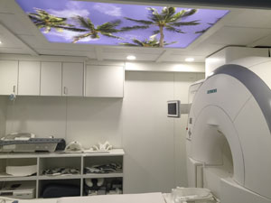
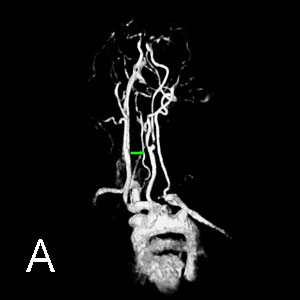
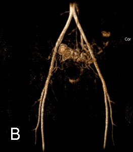
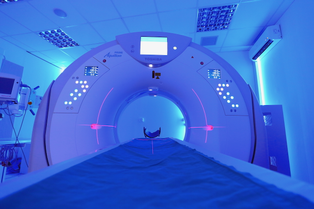
Certain imaging may also require contrast media to provide a better presentation of the pathological process and to provide a more accurate diagnosis. The modern 1.5T, which is also available in our institution (Figure 1, Siemens Essenza in our institution), in combination with the latest powerful software, enables continuous contrast imaging even in situations where this was not possible before (Figure A. Contrastless representation of neck blood vessels and brain, Fig. B. Contrastless imaging of the pelvic and leg arteries).
Anesthesia MRI
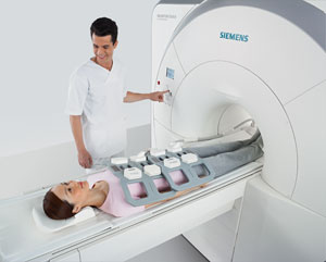
Where necessary, usually for children under the age of 5 and restless or claustrophobic patients, brief anesthesia may be given to obtain quality recordings for diagnosis. Anesthesia is performed in collaboration and under the supervision of an anesthesiologist.
Important recording note!
A doctor or engineer of honey is necessary before recording. radiology report on:
"pacemakers" or other built-in electronic appliances, because of the malfunctioning of these devices in the magnetic field, it is often not possible to record MR (the new generation of pacemakers, stents can go for MRI imaging)
metal bodies in the body (bullets, debris, buckles after surgery on blood vessels, metal plates ...)
Pregnancy: The harmful effects of MR imaging alone have not been proven, the contrast containing metal gadolinium crosses the placenta into the fetus and is suspected to be damaging. Therefore, women who may be pregnant should mention this before recording.
breastfeeding is not a contraindication for MR examination, but if contrast is used, stopping breastfeeding for the next 24h is advised.
No objects of magnetic material (credit cards, cell phones, keys,…) should be brought into the room with the appliance…
dental implants and fillings are a contraindication for this review!
There are two basic types of magnetic resonance imaging equipment. The open ones use permanent magnets and the closed ones use a magnetic field that is generated by electricity. Closed-loop appliances are of a stronger field, generally 0.5 to 3 T (Tesla) and take shorter inspections.
By magnetic field strength, MR imaging devices are divided into:
low field devices - up to 0.5 T
medium field devices - 0.5 T to 1 T
high field devices - 1 T or more (1.5 T, 2 T, 3 T, 7 T,…)
The quality of the display is proportional to the strength of the magnetic field, but this is not the only factor - the quality of the image also depends to a large extent on the image processing software used and the knowledge of the engineer who takes the image.
MRI examinations
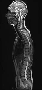
Magnetic resonance imaging provides extremely accurate analyzes of soft tissue structures such as the brain, spinal cord, abdominal organs, small pelvis, lungs, which were previously unavailable, as well as realistic anatomical representation of a particular region and localization of the lesion or foci in it, like no other technique. views (Figure 5.6.7).
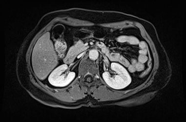
Abdominal MRI
Unlike CT, there is no radiation (especially significant for younger people, pregnant women, certain patients) and has excellent contrast resolution of certain tissue lesions, tumors, etc. It is especially important that these methods can be seen at the stage when they are small in size , and cannot be recognized by other diagnostic procedures (Uz, CT).
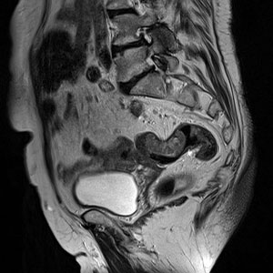
MRI of the small pelvis
It shows the condition of the rectum, rectosigmoid part of the colon, vagina, uterus, ovary, fallopian tube, bladder, prostate, seminal vesicles, regional lymph nodes. MRCP This examination shows the gallbladder, bile ducts and successfully replaces the classic, invasive ERCP examination. Precise insight into the condition of the gallbladder and biliary tract is obtained, and the existence of calculus, etc. is successfully detected. damage up to 3 mm in size.
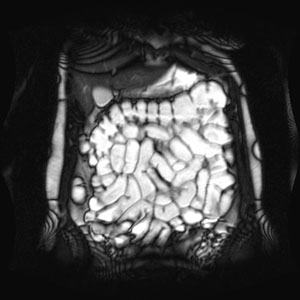
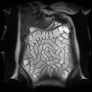
MRI colonography
It is a method of screening the colon (if necessary, thin) with magnetic resonance, which requires a stronger field apparatus and with the latest imaging software, and is an elegant and comfortable alternative to classical colonoscopy or MSCT colonography as an excellent screening method, completely painless, inflatable , no stripping, no discomfort, no radiation.Preparation is like for a classic coloscopy, and the recording itself does not require removal, inflating,…
MRI of the spine, in the diagnosis of neck and back pain
For neck and back pain, MRI is the best diagnostic method!
They see very well:
spinal column anomalies
spinal cord injuries
inflammatory processes on vertebrae and spinal cord
hernia discus
tumors of the vertebrae, nerves and spinal cord
diseases affecting the bone marrow
cysts (arachnoid, perineural, dermoid, cysts of the spinal joints).
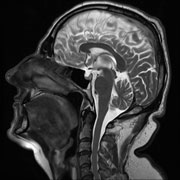
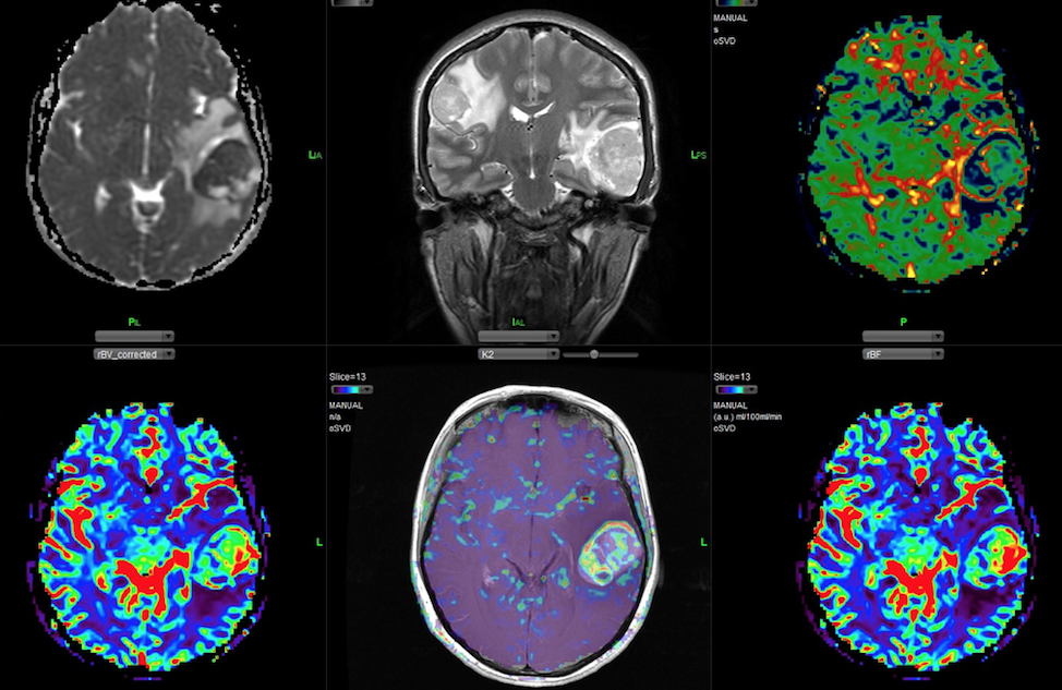
Brain MRI
MRI in the diagnosis of headache
On the other hand, magnetic resonance imaging is an indispensable tool for the diagnosis of secondary headaches, more specifically the diseases that cause them. The causes of secondary headaches are therefore other, sometimes serious, illnesses, injuries or tumors.
MRI examination can also diagnose headaches resulting from the contact of blood vessels with cranial nerves. This is most often the case for the fifth neuralgia (trigeminal neuralgia).
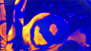
MRI of the heart and blood vessels
MRI is an excellent method in the diagnosis of some diseases and problems of the heart and blood vessels (especially in:
heart defects
thrombus, heart tumors
diagnosis of myocarditis
analysis of viable myocardium, contractility, scarring, perfusion of heart muscle of valve disease
aortic diseases-aortography
diseases of the blood vessels of the neck and head arteries
arteriography
peripheral artery disease
veno-venography diseases
diseases, primarily narrowing of the renal arteries (renography)
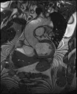
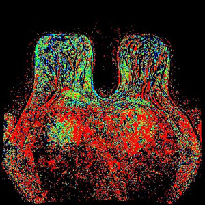
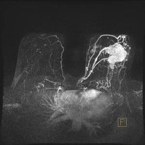
Breast MRI
Breast MRI is an excellent method of showing changes in the breast, which completes the diagnosis of breast disease, which includes ultrasound, mammography, elastography, FNAC, core biopsies
MRI of joints, bones, muscles, musculoskeletal structures
The method is precise and for the time being, above all diagnostic methods, especially for the presentation of ligaments within the joints.